Optic Atrophy
Optic Atrophy
Optic atrophy is a pathological term referring to optic nerve shrinkage caused by the degeneration of retinal ganglion cell (RGC) axons. The term “optic atrophy” is regarded as a misnomer since atrophy implies disuse.
Therefore, a better term for optic atrophy would be “optic neuropathy.” However, that term is also controversial since, in certain situations, such as primary optic atrophy or traumatic brain injury, optic neuropathy may not occur.
Optic atrophy is the end stage of a disease process affecting the retinogeniculate portion of the visual pathway, characterized by a non-specific sign of optic disc pallor. While the peripheral nervous system has an intrinsic ability for repair and regeneration, the central nervous system, for the most part, is incapable of such processes. The axons of the optic nerve are heavily myelinated by oligodendrocytes and reactive astrocytes, which express many inhibitory factors for axonal regeneration.
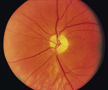
When light is thrown on the fundus from a light source, it undergoes total internal reflection through the axonal fibers. Subsequently, reflection from the capillaries on the disc surface gives rise to the characteristic yellow-pink color of a healthy optic disc. In eyes with cataract, red color is exaggerated, giving rise to a hyperemic appearance of the disc. Conversely, in pseudophakic individuals, the disc may appear to have some degree of pallor.